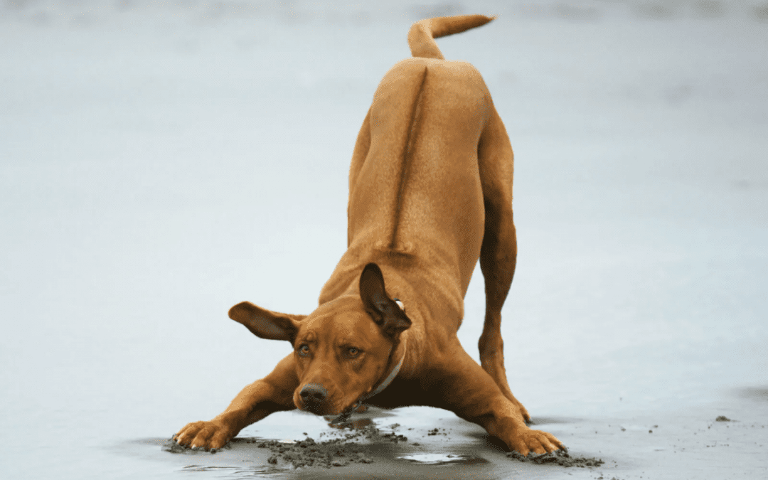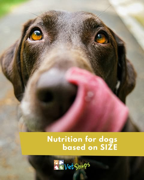
Nutrition Guide For Dogs, Based On Your Pet’s Size
When it comes to feeding dogs, their size plays a crucial role in determining their nutritional needs.
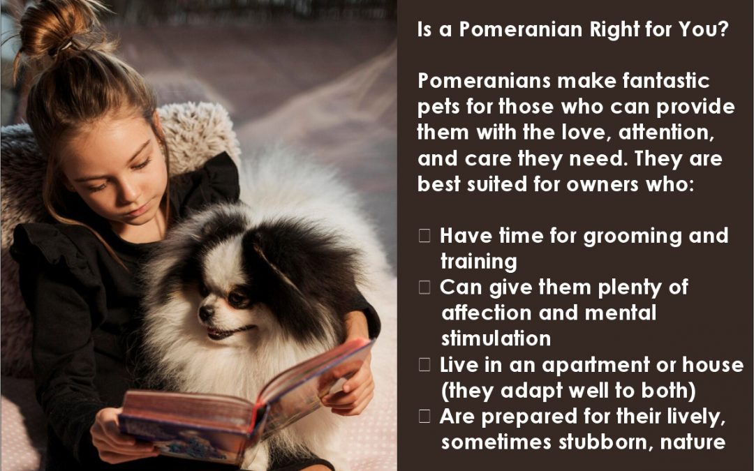
Small breeds, such as Chihuahuas and Pomeranians, typically require a diet that is higher in calories and protein relative to their body weight, as they have faster metabolisms and need more energy to sustain their activity levels.
On the other hand, medium-sized dogs like Beagles and Bulldogs benefit from a balanced diet that includes a mix of proteins, fats, and carbohydrates to support their moderate energy levels and overall health. Large breeds, such as Golden Retrievers and Great Danes, require a diet that is lower in calories but rich in essential nutrients to promote healthy growth and prevent obesity, which can lead to joint issues. Additionally, it’s important to consider the life stage of the dog—puppies, adults, and seniors all have different dietary requirements that should be addressed to ensure optimal health and longevity.
By tailoring a dog’s nutrition to their size and life stage, owners can help their furry friends thrive and maintain a healthy weight.
#petsupplies #petcare #shoplocal #petlovers #petproducts #smallpets #animalcare

Mini Adult Dogs

Small Breeds (1-10kg)

Medium Breeds (11-25kg)

Large Breeds (26-44kg)

Giant Breeds (45kg+)
Whilst you are here, Shop some Products for your DOG;
Showing 1–12 of 164 resultsSorted by popularity
-

Adult Yorkshire Terrier Pouch 85g, Dogs Food, KimVet e-Shop, Royal Canin
R24.16 -
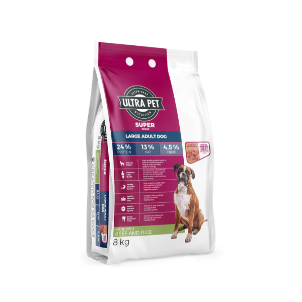
Adult Large Superwoof Beef, Dogs Food, KimVet e-Shop, Ultra Pet
R600.00 – R2,345.01 -
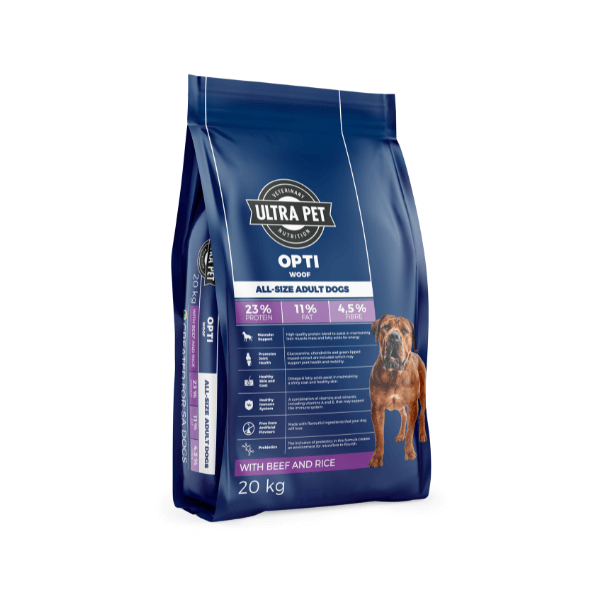
Adult Optiwoof, Dogs Food, KimVet e-Shop, Ultra Pet
R449.01 – R1,829.01 -
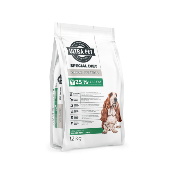
Adult Weight Control Special Diet, Dogs Food, KimVet e-Shop, Royal Canin
R215.01 – R760.01 -
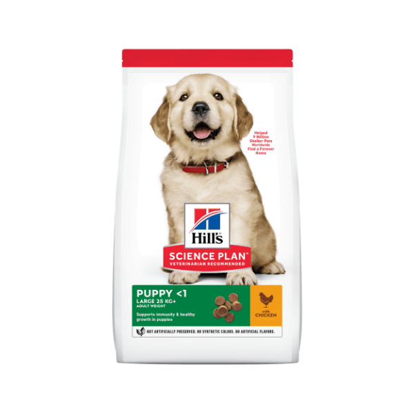
Puppy Large, Dogs Food, KimVet e-Shop, Hill’s Science Plan
R429.01 – R1,708.99 -

Adult Large Superwoof Chicken, Dogs Food, KimVet e-Shop, Ultra Pet
R600.00 – R2,345.01 -
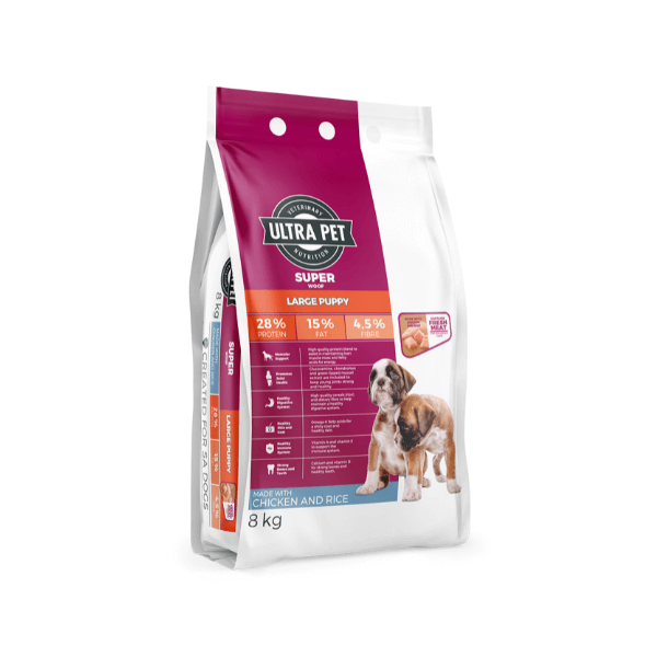
Puppy Large Superwoof, Dogs Food, KimVet e-Shop, Ultra Pet
R245.00 – R1,375.01 -
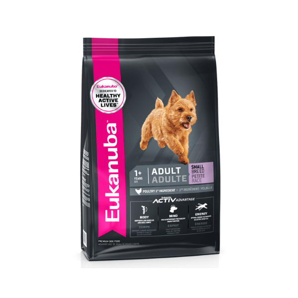
Adult Small, Dogs Food, KimVet e-Shop, Eukanuba
R131.53 – R1,588.11 -

Adult Maintenance, Dogs Food, KimVet e-Shop, Nutribyte
R107.79 – R1,698.22 -

Adult Sensitive Stomach & Skin Medium & Large, Dogs Food, KimVet e-Shop, Hill’s Science Plan
R465.00 – R1,615.00 -

Adult Senior Vitality Small & Mini, Dogs Food, KimVet e-Shop, Hill’s Science Plan
R275.01 – R999.00 -
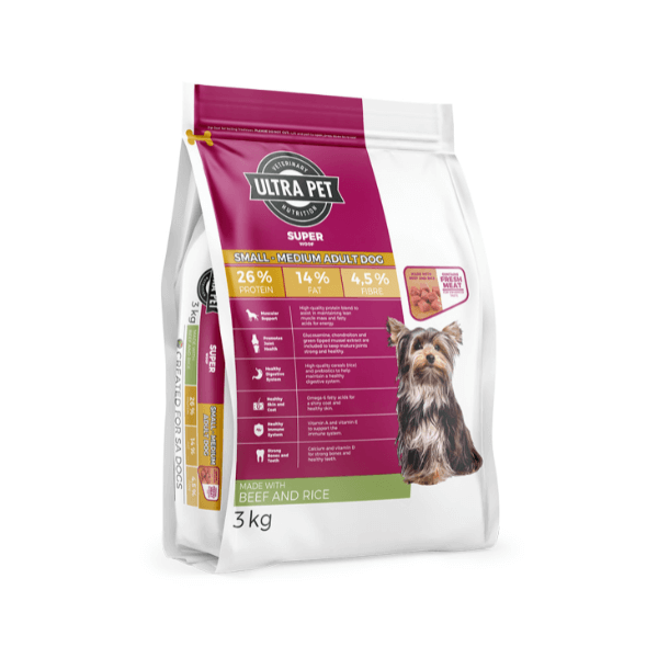
Adult Small – Medium Superwoof Beef, Dogs Food, KimVet e-Shop, Ultra Pet
R265.00 – R600.00